Watching a loved one navigate the recovery process after a major operation can be a source of significant stress for many families․ Managing drains and tubes after surgery is a delicate task that requires precision, cleanliness, and a vigilant eye for subtle changes in health status․ We understand that the presence of medical equipment at home may feel overwhelming, but with the right guidance, it becomes a manageable part of the healing journey․ Our commitment is to ensure that every family feels supported and informed as they maintain the clinical purity necessary for a safe and infection-free recovery․
Clinical Quick Answer
Properly managing drains and tubes after surgery involves maintaining a sterile environment, measuring output accurately, and ensuring the collection devices maintain their suction․ Engaging professional Nurse Services can significantly reduce the risk of surgical site infections and post-operative complications by providing expert oversight at home․ Clinicians focus on monitoring the color and volume of drainage to ensure the body is healing correctly while preventing fluid accumulation that could lead to pain or abscess․
Understanding the Vital Role of Post-Surgical Drains
Managing drains and tubes after surgery is not merely a technical chore; it is a fundamental component of physiological stabilization․ After an invasive procedure, the body naturally produces inflammatory fluids, including blood and serum, which can collect at the surgical site․ If these fluids are not effectively evacuated, they can create a pocket known as a seroma or a hematoma․ These pockets not only cause significant pressure and pain but also serve as a breeding ground for bacteria, potentially leading to deep-tissue infections․
- Prevention of Tissue Tension: By removing excess fluid, drains reduce internal pressure, allowing the surgical edges to approximate and heal more efficiently․
- Infection Control: Removing stagnant fluid eliminates the medium where pathogens thrive, lowering the risk of post-operative sepsis or localized abscesses․
- Diagnostic Value: The characteristics of the drainage-such as its consistency and color-provide immediate feedback to the medical team about internal healing or potential hidden bleeding․
- Facilitating Mobility: When fluid is properly managed, patients often experience less localized swelling, making it easier for them to participate in physical therapy and early ambulation․
Common Types of Drains and Tubes Encountered at Home
When families take on the responsibility of managing drains and tubes after surgery, they may encounter several different devices, each with its own mechanical principles․ Understanding whether a drain is active (suction-based) or passive (gravity-based) is crucial for correct maintenance․ Nurses often educate caregivers on these distinctions to ensure the equipment functions as intended by the surgeon․
- Jackson-Pratt (JP) Drains: These consist of a thin, flexible tube connected to a bulb-shaped reservoir․ They rely on negative pressure created by squeezing the bulb to pull fluid from the wound․
- Hemovac Drains: Similar to JP drains but typically larger and circular, Hemovacs use a spring-loaded mechanism to create suction, often used in orthopedic or large-incision surgeries․
- Penrose Drains: These are flat, latex or silicone ribbons that act as passive drains, allowing fluid to flow out onto a dressing via capillary action and gravity․
- Pigtail Catheters: Often used for internal abscesses or pleural effusions, these tubes are coiled at the end to stay in place and usually drain into a bag․
- Biliary or T-Tubes: Specifically used after gallbladder or liver surgeries to drain bile and prevent a buildup that could cause jaundice or chemical peritonitis․
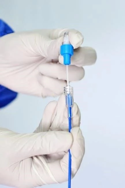
Sterile Technique and Daily Maintenance Protocols
Maintaining clinical purity while managing drains and tubes after surgery requires a strict adherence to aseptic techniques․ The entry point of the tube is a direct portal into the body, making it highly susceptible to contamination․ Professional caregivers emphasize that every interaction with the drain must begin with thorough hand hygiene and the use of clean, medical-grade gloves․
- Handwashing Standards: Caregivers should wash hands with antimicrobial soap for at least 20 seconds before and after touching any part of the drainage system․
- Site Cleaning: The skin around the insertion site should be cleaned daily using sterile saline or as directed by the surgeon, typically wiping in a circular motion from the center outward․
- Securing the Device: Drains should be pinned to the patient’s clothing (not the skin) to prevent accidental pulling or dislodgement during movement․
- Maintaining Suction: For bulb drains, the reservoir must remain compressed․ If the bulb is full or inflated, it is no longer providing the negative pressure needed for fluid removal․
- Emptying Frequency: Drains are generally emptied every 8 to 12 hours, or whenever they become half full, to maintain consistent suction levels․
Monitoring Output: Differentiating Normal and Concerning Fluids
One of the most critical aspects of managing drains and tubes after surgery is the accurate documentation of fluid output․ This data is the primary tool used by surgeons to decide when a drain can be safely removed․ The progression of fluid color is a standard clinical indicator of the healing phase the body is currently navigating․
- Sanguineous Phase: In the first 24 hours, drainage is typically bright red (bloody), which is expected immediately following tissue disruption․
- Serosanguineous Phase: As healing begins, the fluid turns pinkish or watery-red․ This transition indicates that active bleeding has slowed․
- Serous Phase: The final stage of drainage is a clear or straw-colored liquid․ This is the goal state and suggests that the internal wound is stabilizing․
- Volume Trends: A steady decrease in the daily volume of fluid is expected․ A sudden, sharp increase in volume may indicate a new internal bleed or a seroma rupture․
- Clot Awareness: Small, occasional clots in the tubing are common, but large amounts of clotted material can block the tube, requiring “milking” or professional intervention․
Identifying and Preventing Complications
Vigilance is the cornerstone of managing drains and tubes after surgery at home․ Complications can arise quickly, and early detection often prevents the need for hospital readmission․ Families must be educated on “red flag” symptoms that require immediate contact with the surgical team or home health nurse․
- Signs of Infection (Surgical Site Infection): Look for spreading redness (cellulitis), increased warmth around the site, foul-smelling drainage, or purulent (pus-like) fluid․
- Loss of Patency: If the drain stops producing fluid suddenly but the area around the incision becomes swollen and painful, the tube may be kinked or blocked internally․
- Skin Integrity: Constant moisture or leakage around the tube insertion site can cause skin maceration and breakdown, which requires barrier creams or specialized dressings․
- Systemic Symptoms: A fever over 101․5°F (38․6°C), chills, or sudden lethargy can indicate that a localized infection has become systemic․
- Dislodgement: If the tube is accidentally pulled out, it should never be pushed back in․ The site should be covered with a sterile dressing and the doctor notified immediately․
The Integration of Professional Nurse Services in Recovery
For many families, the transition from hospital to home is made safer through the utilization of professional nursing support․ Experts specializing in managing drains and tubes after surgery bring a level of clinical oversight that reduces the burden on family caregivers․ Nurses not only perform the technical tasks but also provide the necessary education to ensure the home environment remains a sterile healing space․
- Expert Assessment: Nurses can distinguish between normal post-surgical inflammation and the early stages of a complication that a layperson might miss․
- Streamlined Communication: Home health nurses act as a bridge, reporting daily output and site conditions directly to the surgeon, ensuring that interventions happen in real-time․
- Patient Comfort: Professionals are skilled in “milking” tubes to clear obstructions without causing trauma to the tissue, a task that many families find intimidating․
- Emotional Support: Having a professional present reduces the anxiety of the patient and family, fostering a more positive psychological environment for recovery․
- Removal Coordination: Nurses often perform the final drain removal in the home setting or coordinate the timing of the final office visit based on clinical data․
Nurse Insight: In my experience, the most common mistake families make is forgetting to “prime” the suction bulb after emptying it․ When managing drains and tubes after surgery, the drain is only effective if the bulb is squeezed tight before the cap is replaced․ I always tell my patients to think of it like a vacuum; without that squeezed bulb, the fluid just sits inside the body․ Also, keeping a dedicated logbook on the nightstand-not just for volume, but for the color and the time of day-makes the surgeon’s follow-up visit much more productive and often leads to the drains being removed sooner․
Frequently Asked Questions
How do I know if the drain is working properly?
Can I take a shower with a surgical drain still in?
What should I do if the fluid looks cloudy or smells bad?
Is it normal to have some leakage around the tube insertion site?
When are drains usually removed?
Contact ProLife Home Care NYC for a free clinical assessment:(718) 232 – 2777
Contact ProLife Home Care NYC for a free clinical assessment: (718) 232-2777